While serotonin is most well known as an essential brain neurotransmitter, it has been estimated that as much as 95 percent of the body’s serotonin is produced in the digestive tract. Similarly, the hormone melatonin is most well known as being produced by the pineal gland at night based on circadian rhythm factors. However, it’s been observed that the digestive tract contains at least 400 times more melatonin than the pineal gland while having no circadian based secretion cycle. It is believed that melatonin levels in the gut are independent of pineal production since rats with pinealactomy were not effected in terms of their gut melatonin concentrations.
In 2005, the Scandinavian Journal of Clinical and Laboratory Investigation published a new discovery (back then) of large amounts of bufotenine (5-HO-DMT) in the stool samples of humans. The levels of bufotenine in stool samples exceeded the amount in urine levels by 1000%. Bufotenine is one of the 3 DMT(s) found within humans (the others being N,N-DMT & 5-MEO-DMT). The researchers proposed that glandular epithelial cells of the small and large intestine are possibly the source of bufotenine in the stool.
While much of the discussion regarding endogenous DMT(s) (as well as melatonin) revolves around the pineal gland, it appears as though the digestive tract has the potentiality to be an overlooked source of DMT(s) production. The question is… where exactly might melatonin, serotonin, and possibly DMT(s) be produced in the gut?
Glial cells are non-neuronal cells in the central nervous system (CNS) and the peripheral nervous system (PNS). In the CNS, glial cells have a wide array of functions including the hormonal regulation of synaptic function/plasticity, myelin formation, cognition, sleep, and the response of nervous tissue to injury. The different types of glial cells within the CNS are as follows: astrocytes, oligodendrocytes, ependymal cells and microglia.
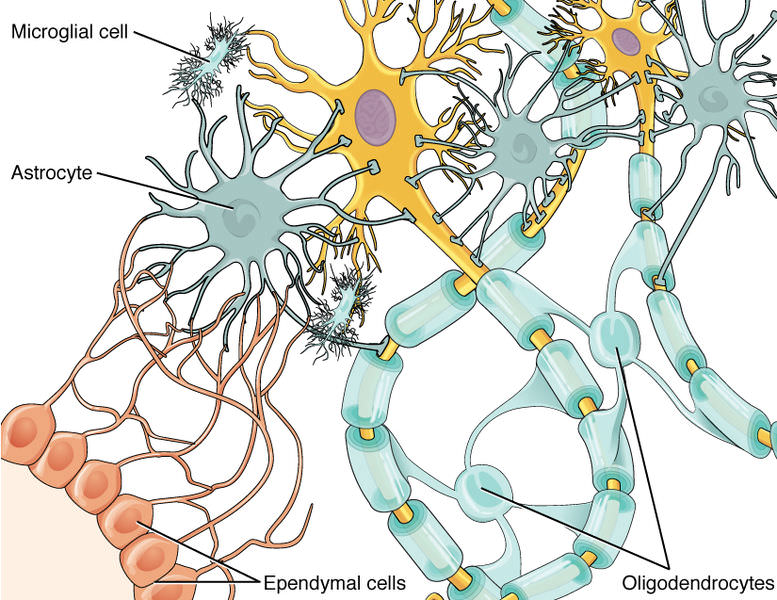
There are also two types of glial cells in the peripheral nervous system (PNS) known as Schwann cells and satellite cells. For simplification purposes, we shall focus on the CNS and specifically the glial cell type known as astrocytes.
Astrocytes are the most abundant type of glial cell in the brain with the primary function of regulating the transmission of electrical impulses. They also play a role in the secretion and absorption of neurotransmitters and maintenance of the blood–brain barrier. A 2007 in vitro study in the Journal of Pineal Research observed for the first time that astrocytes from the rat cortex and glioma C6 cell line synthesized melatonin. The study also found the presence of serotonin and the two key enzymes in the pathway of melatonin synthesis (N-acetyltransferase & hydroxyndole-O-methyltransferase) in the cultured astrocytes. Release of melatonin into the culture medium showed no diurnal changes. A 2010 study in the Journal of Neurological Sciences also found melatonin and it’s precursors in the astrocytes of the gerbil hippocampus. These findings indicate that brain melatonin levels are modulated by astrocyte synthesis and secretion. The percentage level of total modulation in comparison to pineal excretion would be very difficult to quantify based on the sheer number of astrocytes (estimated to be in the billions) throughout the brain as well as the challenges of verifying the amount of secretion per each astrocyte in vivo in living humans. Nevertheless, astrocyte melatonin secretion might offer a partial answer as to why pinealectomy (removal of the pineal gland) fails to completely disrupt the circadian rhythm. However, there is a likely correlation between lesser neural melatonin levels and reduced theta wave amplitude during REM sleep of pinealectomized rats compared to controls. This is based on the findings that exogenous melatonin administration amplifies theta wave strength.
*It should be noted that melatonin has been shown to be synthesized in the following areas of the body (humans & various animals) using the same pathway as the pineal gland & gut: retina, ciliary body, lens, harderian gland, brain, thymus, airway epithelium, bone marrow, ovary, testis, placenta, uterus, lymphocytes, mast cells, skin, neurons and platelets.
Scientists have developed the name “enteric glial cells” (EGCs) to describe intestinal glial cells that have been described as being extremely similar to astrocytes in terms of morphological characteristics and expression markers. A book titled “Enteric Glia” published in 2014 by Dr. Brian Gulbransen describes the many functions of EGCs, their vast similarities to astrocytes, as well as the minor differences between the two types of cells. His summary regarding the differences is as follows: “The two cell types undoubtedly display many similarities but specific differences indicate that enteric glia are fundamentally different from astrocytes. First and foremost is that fact that enteric glia and astrocytes have different developmental origins: enteric glia are derived from the neural crest while astrocytes are derived from precursor cells that line the neural tube. This difference is highlighted by findings demonstrating that enteric glial cell development requires neuregulin signaling through the ErbB3 receptor while astrocytic development does not (Riethmacher et al., 1997). Further, mature enteric glia lack of expression of the astrocytic marker aldehyde dehydrogenase 1 family member L1 (Aldh1L1) (Boesmans et al., 2014). Therefore, generalizing astrocytic properties to enteric glia for the purpose of modeling or developing hypotheses is a useful principle but one must keep in mind the unique nature of enteric glia.”
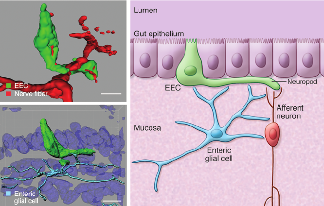
To complicate matters further, a 2017 study in the journal Scientific Reports observed that “astrocyte-like” glial cells were generated from neural crest cells in the developing spleen of birds and primates. Not to digress in extreme fashion but if the main differentiation between EGCs and astrocytes are origination (not functionality)… and astrocytes have been observed to originate from the same origin (neural crest cells) as EGCs, then the clear cut differences begin to wane. I understand the need to incessantly find new labels for “things” but perhaps EGC’s can be referred to as gastrocytes (short for gastrointestinal astrocytes) or something of that nature being that for all intensive purposes it appears as though EGC’s have virtually identical function to astrocytes? Other factors to take into account are “epi” factors in which microenvironments alter gene expression such as the cited astrocytic marker (Aldh1L1). It should be quite obvious that the environment of astrocytes in the brain and gut are quite different leading to localized gene expression changes. A less dramatic example of “epi” factors are cited in a 2011 study in the journal Glia which observed decreased ALDH1L1 expression levels in the spinal cord during postnatal maturation while expression was up-regulated in reactive astrocytes in both acute neural injury and chronic neurodegenerative conditions. A 2014 study in Neuroscience Letters found that ALDH1L1 was in fact expressed in EGCs outside of the myenteric ganglia hence the title of the paper, “The astrocyte marker ALDH1L1 does not reliably label enteric glial cells”. It doesn’t seem as though there is a definitive differentiation between EGC’s and astrocytes from our perspective.
Apologies for the digressive rant…
Back to the topic at hand.
We believe that much like the astrocytes in the brain have been observed to synthesize melatonin, astrocytes (EGCs if you must) throughout the digestive tract are a potential source of serotonin, melatonin, and bufotenin. Perhaps the researchers from the 2005 bufotenin study are referring to astrocytes/EGCs when hypothesizing that stool bufotenin originates from intestinal epithelial cell secretion?
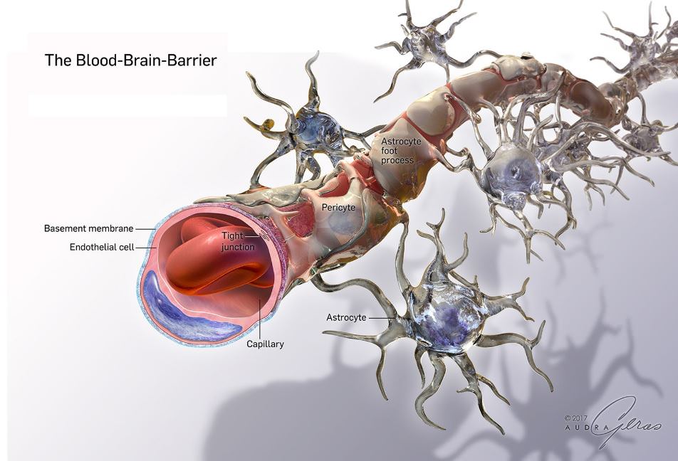
A 2010 review in Acta Neuropathologica titled “Astrocytes: Biology and Pathology” covers numerous studies that observe a distinct relationship between astrocytes and epithelial cells in the brain and gut. In vitro experiments suggest that astrocytes and related glial cells induce barrier properties at epithelial layers leading to key roles in the blood brain barrierfunction as well as gut barrier function. These barriers (gut/brain) allow compounds related to proper function to permeate the system while also serving as a protective safeguard against pathogens. It’s been cited that inflammation allows for greater permeability of both the gut and brain barriers. Nevertheless, further studies will be needed to verify that bufotenin is produced in gastrointestinal astrocytes (EGCs if you must).
Since functionality is always of immense focus at DMT Quest, the natural question one must ask is… why is a “hallucinogen” such as bufotenin produced in the GI tract in the first place?
Thus far, it appears as though both N,N-DMT and 5-MEO-DMT act as endogenous anti-inflammatory, anti-oxidative agents in addition to their “hallucinogenic” properties. Being that bufotenin has significant overlap with these DMT(s) in terms of receptor affinity, molecular structure, chemical/enzymatic precursors, and hallucinogenic properties, we believe that there is a high probability that bufotenin also acts as an anti-inflammatory/anti-oxidative agent. A 2013 review in BioMed Research International discusses the link between oxidative stress, colorectal cancer, and the various factors that effect the ability of the gut epithelial cells to cope with metabolic challenges. We believe that bufotenin is one of the numerous endogenous anti-inflammatory compounds that aids in increasing the integrity of the gut barrier. A 2016 review in the journal Free Radical Biology and Medicine highlights the mechanisms of adapative regulation of the brain’s antioxidant defenses with a focus on astrocytes.
INMT
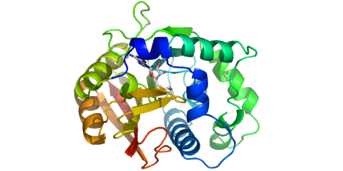
Indolethylamine N-methyltransferase (INMT) is an enzyme in the body that converts tryptamine to N,N-DMT and serotonin to bufotenin. One of the most abundant concentrations of INMT in the mammalian body is the lungs (possibly related to the abundance of epithelial cells lining the alveoli). An excellent review of INMT’s functions in relationship to endogenous DMT(s) was published in 2018 in the journal Frontiers in Neuroscience by Jon G. Dean.
A related interesting excerpt from the 2005 bufotenin study is the following: “The enzyme indolethylamine N-methyltransferase (INMT), which is capable of forming DMIAs (DMT(s)) and is present in several mammalian tissues, has recently been cloned from rabbit and human lung. It is present in the majority of stromal and epithelial cells of organs related to the autonomous nervous system, but absent from neurons and striated muscle cells (Karkkainen et al., unpublished observations, 2004).”
DMT Quest would reach out to Dr. Karkkainen regarding the reasons for the unpublished observations. He stated, “The study you are referring to was never published. We started collaboration with Professor Weinshilboum of Mayo Clinic after having read the two studies of INMT of rabbit and human lung. In this work the Mayo clinic was responsible for making histochemistry and myself for writing the publication etc. I even wrote a prelimininary manuscript. I retired (old age) 15 years ago and the INMT group in Mayo clinic decomposed.”
We find it intriguing that it was observed that INMT was present in the stromal and epithelial cells of organs related to the autonomous nervous system being that much of the discussion at DMT Quest relates to generating conscious control over this system. Stromal cells are connective tissue cells of any organ that support the function of the parenchymal cells (functional parts) of that organ. A 2018 write-up in the Proceedings of the National Academy of Science notes that the stroma cells of the gut include fibroblasts, myofibroblasts, fibrocytes, hematopoietic cells, neural, and enteric glial cells (EGCs). As we’ve outlined above, the functional characteristics of EGCs and astrocytes appear virtually identical with the main difference being anatomical origination. If EGCs do in fact contain INMT, that furthers the case for bufotenin secretion being that the biochemical precursor (serotonin) and enzyme (INMT) are present at the site. A 2015 in vivo study in the American Society for Neurochemistry found a protein secretion normally attributed to astrocytes/EGCs within mouse lungs (lung glial cells are referred to as non-myelinating Schwann cells). A 2009 study in the journal Laboratory Investigation identified stromal cells in human lung tissue. Perhaps it would behoove us to succinctly identify which type of cell(s) in the lungs are providing the INMT as it could assist us in comprehending other region specific mechanisms for DMT(s) synthesis?
Epithelial cells line the outside (skin) and the inside cavities of the body including the insides of the lungs, reproductive tract, and urinary tract. In the academic book Biology: Concepts and Applications, it cites the following: “endocrine glands comprise of clusters of hormone-secreting epithelial cells embedded into connective tissue and well supplied with blood vessels.” A 2001 study in the journal Glia found that epithelial cells from the choroid plexus of mice differentiated into astrocytes following implantation into the spinal cord. While it appears clear that there is an intimate relationship between epithelial cells and astrocytes, there is no data (thus far) to support the notion that astrocyte/EGC INMT is the direct source of epithelial INMT throughout the autonomic nervous system.
*There lies the potentiality that the studies purporting to find little to no INMT in the brain need to be re-evaluated for accuracy and efficacy. It appears as though a 1971 study published in Nature found INMT in the brains of humans and sheep. The study found the highest values of INMT in the uncus, medulla, and amygdala regions of the brain. Interestingly enough, the medulla and amygdala have both been found to contain significant concentrations of astrocytes. Perhaps the more recent studies claiming to find no INMT concentration in the brain inadvertently compromised the integrity of the neural sampling by grinding up the whole brain tissues prior to testing for INMT? It would seem important to verify the truth of INMT levels and locations in the brain being that the discussion is so pertinent to endogenous DMT(s) synthesis. It seems obvious at this point that a “neuron-centric” focus of INMT in the brain would be an unlikely source while surrounding glial cells offer a more likely alternative.
Receptors
Much of the discussion regarding DMT(s) effects in the body revolves around receptor affinity. Thus far much of the focus has pertained to the 5-HT2A and Sigma-1 receptors.
A 1999 study in the journal Experimental Neurology observed that human brain tissues from patients suffering from various forms of neurological damage (Alzheimer’s, hypertensive encephalopathy, cerebral infraction, etc.) experienced an activation of astrocytes. The activated astrocytes showcased strong 5-HT2A (but not 5-HT2B or 5-HT2C) and glial fibrillary acidic protein (GFAP) receptor immunoreactivity. The researchers suggest that the upregulation of 5-HT2A receptors potentially signify an early response reaction in astrocytes designed to maintain homeostasis.
A 2018 review in the journal Advances in Experimental Medicine and Biology outlines the role of Sigma-1 receptors in neurodegeneration as well as neuroregeneration. It cites numerous studies in which Sigma-1 receptors have been identified in astrocytes. A 2016 study in the journal Neuropharmacology found that spinal cord injuries activate the expression of Sigma-1 receptors in astrocytes.
As we’ve touched upon in the past in pieces such as “Cancer & DMT” and “Revisiting the Schizophrenia-DMT Relationship”, we believe that upregulation of endogenous DMT(s) is the body’s attempt to maintain homeostasis and induce repair. The upregulation of receptors within astrocytes in which DMT is known to have affinity for is not surprising especially in regards to inducing repair mechanisms. Neuroplastic effects of the shamanic brew Ayahuasca have been observed much like the neuroplastic effects of meditation.
A 2017 study in the journal Nature Neuroscience observed that glial cells and not neurons lead the way in terms of brain assembly in worms. Being that astrocytes are the most abundant glial cell in the brain, we assume that they are playing an integral part of brain assembly. The researchers state that there is enough similarity between worm brain development and that of vertebrates to note the significance of this finding and the implications for our understanding of neurobiology. A 2016 review in the journal Frontiers in Human Neuroscience outlines the many essential roles that astrocytes and microglia play in human brain development.
Not to be relegated solely to discussing brain development, in Dr. Robert O. Becker’s book “The Body Electric”, he documents a study in which he was attempting to find the driving forces behind limb regeneration and healing of fractures. Here is the excerpt:
“After Bruce had worked out the complicated surgical procedure, we anesthetized a series of rats, removed the nerve supply to one leg of each animal, and broke the fibula, or smaller bone of the calf, in a standard way. Then every day we reanesthetized a few of the rats and took out the fracture area to mount it for the microscope. At the same time, Bruce checked the cut nerves to make sure there was no regrowth. Successful denervation was confirmed by the microscope and by complete paralysis of the affected leg.
The results were encouraging yet puzzling. The nerves didn’t regrow, and the broken bones took twice the normal six or seven days to heal, but heal they did, even though theoretically they shouldn’t have knit at all without nerves.
It was well known that the severed end of a nerve would die after a couple of days, but, since we’d cut the nerves at the same time as we’d broken the bones, maybe the cut ends had exerted a subdued healing effect while they remained alive. In another series of animals, we cut the nerves first. Three days later, after making sure the legs were fully denervated, we operated again to make the fractures. We felt sure the delay would give us true nonunions. To our surprise, however, the bones healed faster than they had in our first series, although they still took a few days longer than normal.
Here was a first-class enigma. The only thing we could think of doing was to cut the nerves even earlier, six days before the fractures. When we got that series of slides back, we found that these animals, whose legs were still completely without nerves, healed the breaks just as fast and just as well as the normal control animals. Then we took a more detailed microscopic look at the specimens Bruce had taken from around the nerve cut. We found that the Schwann cell sheaths were growing across the gap during the six-day delay. As soon as the perineural sleeve was mended, the bones began to heal normally, indicating that at least the healing, or output, signal was being carried by the sheath rather than the nerve itself. The cells that biologists had considered merely insulation turned out to be the real wires.”
While Schwann cells are not astrocytes, they are a type of glial cell in the periphery nervous system. Interestingly enough, a 2003 study in the Journal of Neurocytology found 5-HT2A receptor protein and mRNA in the sciatic nerves and Schwann cell cultures of rats. The researchers hypothesize that these 5-HT receptors in Schwann cells are activated by the release of 5-HT from neighboring mast cells (which also happen to secrete melatonin). In addition, a 2004 study in the journal Brain Research identified Sigma-1 receptors within the sciatic nerves and Schwann cell cultures of rats. While the primary focus of this piece is obviously astrocytes, it appears as though there lies the possibility that DMT(s) synthesis and release could be glial-wide throughout the body. Being that in Becker’s study the nerve cells were verified to not be the drivers of limb regeneration/repair and signaling, perhaps a much more extensive dive into astrocytes and the glial system is in store in the coming decades. Much like the field of genetics is beginning to change with the emergence of epigenetic research, the field of neurological research might begin to shift focus from neurons to the surrounding “support” cells that might actually be just as key as the neurons themselves.
Pineal Gland
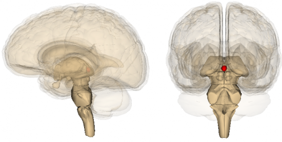
While much of the focus of endogenous DMT(s) discussion (and melatonin for that matter) revolves around the pineal gland, it would seem relevant to discuss the relationship between this gland and astrocytes. The pineal gland itself is comprised of pinealocytes, astrocytes, microglia, and other interstitial cells. In essence, we are not deviating from the pineal discussion entirely as astrocytes are an integral part of pineal anatomy and secretory properties. While the majority of pineal gland cells are comprised of pinealocytes (95%), the relationship between pinealocytes and astrocytes appears significant. A 2013 study in the journal BioMed Research International found that modulation of pineal melatonin synthesis by glutamate involves paracrine interactions between pinealocytes and astrocytes.
In 2013, a study in BioMedical Chromatography would verify the occurrence of all 3 DMT(s), their precursors, and metabolites at the site of the pineal gland in live rats. Melatonin and its precursors were also found in the same study. There were no citations regarding any mention of differentiating between these compounds occurring in pinealocytes or astrocytes.
Based on the information cited throughout this piece regarding astrocytes, INMT, epithelial tissue, and the role of astrocytes at the blood-brain/gut barrier it would seem logical that the brain-DMT(s) discussion would include the choroid plexus. This part of the brain is responsible for producing cerebrospinal fluid (CSF), filtering CSF, and providing a barrier between CSF and peripheral blood. A 2015 review in Frontiers in Neuroscience discusses the data suggesting that the choroid plexus provides “a stem-cell like repository for neuronal and astrocyte glial cell progenitors”. Simply based on the cellular composition of the choroid plexus and the significantly greater levels of N,N-DMT in CSF compared to other endogenous fluids… it would appear that DMT(s) formation should be properly investigated at this site.
Morphogenesis
As we’ve cited in the past, a 2011 frog study in the journal Developmental Dynamics from Tufts University reported the occurrence of bioelectrical signals as being necessary for normal head and facial formation. Patterns of visible bioelectrical signals outlined where the eyes, nose, mouth, and other features later appeared in an embryonic tadpole. Based on the 2017 study showcasing glial cells as being the key drivers in brain development (not neurons) and Dr. Becker’s findings regarding non-neuron based repair capabilities of mammalian limbs, it begs us to bring up the concept of morphogenesis in relation to astrocyte/glial cells.
Being that these occurrences of facial formation, brain development, and limb regeneration/repair are autonomic transpirations, it would appear that the building blocks of autonomic signaling (astrocytes/glial cells) would be important to focus on as the leading biochemical/anatomical correlates to the bioelectrical aspect. There is the possibility that specific bioelectric signaling frequencies upregulate specific matrixxes of biochemicals necessary to physically manifest the signaling. While we discuss increases in gamma wave amplitude in the brain during states potentially associated with endogenous DMT upregulation, there lies the possibility that measurable electrical changes also take place throughout the CNS and PNS.
The next line of questioning pertains to changes in electrical brain activity (consciousness if you will) correlating with changes in neural astrocyte synthesis and secretion of various compounds (not just DMT(s)). What types of secretory changes take place when regions of the brain are stimulated to produce delta waves? alpha waves? gamma waves? coupling of theta-gamma waves? What about fluctuations in neural CO2 levels (pH dependent)? A 2017 study in the Journal of Neuroscience indicates that neural CO2 fluctuations definitively effect astrocyte secretion. Another 2016 study in the Journal of Neuroscience found that brainstem astrocytes detect physiological changes in pH and contribute to the development of adaptive respiratory responses to the increases in the level of blood and brain PCO2 /[H-].
This would be an extremely difficult study to carry out in vivo, in live humans but would be essential to understanding the dynamic changes taking place across the many layers of human physiology. Maintaining a sole focus of understanding biochemical precursors, enzyme driven synthesis, and metabolites is insufficient to comprehending true functionality. Unfortunately, we have many technological hurdles in terms of developing equipment that can reliably offer insights into this type of analysis. Utilizing brain activity/imaging equipment (EEG, MEG, fMRI, DC) while simultaneously taking blood and saliva samples might offer a crude temporary snapshot as to what’s taking place within the body during these altered states. However, being that there’s such a huge discrepancy between the levels of biochemicals at neurological concentrations compared to blood (ex. 2000% difference in nocturnal third venticular CSF melatonin levels compared to blood levels), it hardly offers more than just fodder for more speculative discussions.
An even greater flaw would be attempting to present a correlation between endogenous biochemical release and the measurable levels/effects following exogenous administration. DMT Quest believes that the two have very little in common based on the notion that endogenous DMT(s) is likely released in a site specific manner (astrocytes to neurons?) which would explain why blood levels are virtually undetectable. This is obviously quite different than injecting a person with DMT(s) in their veins and measuring the subsequent effects, levels, and metabolites in the blood. At this point we will have to add some serious footnotes to the piece “Measuring DMT Formation in Humans”.
*In 2017, Dr. Ede Frecska would present at “Breaking Convention” regarding his hypothesis of human physiology and DMT. In this presentation he would state that blood monoamine oxidase (MAO) does not breakdown DMT significantly (citing a 1967 study).
This could be an indicator that DMT(s) are not meant to be in the bloodstream but instead belong at the site of action where they can be utilized by the body in a targeted fashion and either deaminated or recycled properly. Perhaps elevated DMT(s) in the blood coincides with inflammation in which barrier permeability has been comprised?
Many questions…
If we truly wanted to calculate the proposed levels in the brain needed to stimulate a “true DMT experience”, we would have to calculate the total number of astrocytes in the brain (ex. 60 billion glial cells X 20% being astrocytes = 12 billion astrocytes (wild estimate)) multiplied by the secretory rate of one normal functioning astrocyte. Just as an extremely crude example… we could say that each astrocyte (12 billion) secretes 0.1 picogram of DMT (completely arbitrary) per hour equating to 1.2 mg of DMT in the brain. Obviously the number of neural astrocytes and their secretory rate are random figures which could fluctuate based on advances in imaging, changes in brain waves effecting secretion, identifying different types of astrocytes, and measuring the number of astrocytes simultaneously secreting the same compound would be rather difficult to verify. There are also the factors of accumulation and metabolism which needed to be taken into account. Nevertheless, this is what we feel is necessary to understand endogenous DMT(s) synthesis within the body.
*Some researchers have estimated the number of neural glial cells to range as high as 1 trillion. Others in the field have estimated that astrocytes comprise as much as 50 percent of all neural glial cells. As you can see… the random conservative figure given above of 20 percent of 60 billion total glial cells equating to 12 billion astrocytes secreting DMT(s) is quite different than 50 percent of 1 trillion total glial cells (500 billion). A 2016 review in The Journal of Comparative Neurology would question the accuracy of the 1 trillion neural glial cells claim as well as the ratio of neural glial to neurons citing that a new counting method called isotropic fractionator does not support the claim. From a gastrointestinal perspective, a 1981 study in the journal Neuroscience found that the number glial cells outnumbered neurons in the enteric plexuses of rats, guinea pigs, rabbits, cats, and sheep. Yes… many questions still need to be answered.
EEG data
In our “Gamma Wave-series”, we extensively cover what appears to be a correlation between increases in the brain wave known as “gamma” (>30 Hz), a myriad of “mystical states” (meditation, rem sleep, hypnosis, Wim Hof Method, etc.), and a potential correlation with endogenous DMT activation. Exogenous DMT in the form of N,N-DMT, 5-MEO-DMT, and Ayahausca have all been observed to increase gamma wave amplitude when ingested separately.
A 2014 in vitro and in vivo mouse study in the Proceedings of the National Academy of Sciences observed that impaired vesicular release from astrocytes suppressed gamma wave amplitude. This also coincided with a disruption in cognitive function. The researchers would state: “These findings demonstrate, to our knowledge for the first time, that fast neural circuit oscillations (gamma) are tightly regulated by astrocytes. Furthermore, the cognitive and behavioral deficits that we have observed following the disruption of gamma oscillations provide evidence that fast network oscillations are not simply an epiphenomenon of neuronal activity but serve specialized functions that are essential for cognition.”
As we stated in the piece “Aha Moments & Dreaming”, a 2009 study in the journal Current Directions in Psychological Science found that “Aha” moments positively correlated with gamma wave bursts. The following is an excerpt from Brain World magazine regarding the study: “In the volunteers that experienced insight, Kounios and Beeman found a distinctive spark of high gamma activity that would spike one-third of a second before volunteers consciously arrived at an answer. Additionally, the flash of gamma waves stemmed from the brain’s right hemisphere—an area involved in handling associations and assembling parts of a problem. Gamma activity indicates a constellation of neurons binding together for the first time in the brain to create a new neural network pathway. This is the creation of a new idea. Immediately following that gamma spike, the new idea pops into our consciousness, which we identify as the Aha! Moment.”
A 2017 mouse study in the journal Neuron would find that astrocytes play essential signaling roles in inducing new neuronal connections. It was observed that the astrocyte-secreted protein “glypican 4” (Gpc4), plays a key role in pre-synaptic and post-synaptic neurons effectively communicating.
It appears that based on the notion that gamma waves correlate with the formation of new neuronal connections and DMT’(s) relationship (both exogenous and speculatively endogenous) to those occurrences, it further strengthens the notion that astrocytes should be a primary focus for understanding DMT(s) and the brain. These cells secrete hundreds of chemicals (neurotransmitters, neuromodulators, hormones, & metabolic, trophic, plastic factors), contain the pre-cursors and enzymes to synthesize DMT(s), contain 2 key receptors (5-HT2A & Sigma-1), have identified mechanisms with which they directly influence neuronal connectivity, and have an active modulation of gamma wave amplitude points to a good deal of intriguing potentials for further research.
A 2017 mouse study in the journal Frontiers in Neural Circuits observed that astrocytic signaling modulates theta rhythm and REM sleep. While this study was focused on calcium signaling, we believe that astrocyte synthesis of melatonin is potentially involved based on what we’ve covered at DMT Quest regarding melatonin and it’s correlations with increases in slow wave amplitude.
Random Thoughts
In 2016, Dr. Philip G. Haydon would write an article in Cerebrum titled, “The Evolving View of Astrocytes”. In this piece, Dr. Haydon would touch upon the historical evolution of neurological research since the early 1900s in which neuronal activity was the primary focus due to the ability to measure their electrical activity. He cites the fact that astrocytes are “electrically silent” by comparison as the main reason as to why research into this field had been largely ignored. It wasn’t until the 1990s when chemical indicators of biochemical signaling were developed that the activity of astrocytes could be observed. While the neurons speak an electrical language… the astrocytes speak a chemical language.
Perhaps my perceptual naiveness is starting to expose itself but I would presume that since N,N-DMT, 5-MEO-DMT, and bufotenin are all biochemicals… understanding their functionality would require a focus on the layer of the nervous system that utilizes chemical signaling. To focus on neurons without understanding the essential synergy with surrounding physical structures seems overly incomplete.
One aspect of the astrocyte-neuron relationship that needs to be sorted out in the future is in regards to monoamine oxidase (MAO) activity identification. A 2017 review in Frontiers in Pharmacology discusses the data regarding the localization of both MAO-A and MAO-B in the brain. The reviewers discuss the difference in results between in vitro and the examination of an intact brain regarding MAO location. It’s cited that the intact brain of primates and non-primates have shown to primarily express MAO-B in astrocytes while MAO-A is localized primarily in the neuron. However, in vitro studies show conflicting data. Perhaps the DMT(s) are synthesized at the astrocyte (no MAO-A) and quickly broken down as they get dispersed into the neuron (from MAO-A) in order to ensure a stable perceptual field?
A 2009 in vitro study in the Journal of Neural Transmission led by Dr. Nick Cozzi suggests that N,N-DMT has the potential to the achieve high intracellular and vesicular concentrations in neurons based on the SERT and VMAT2 uptake transporters. Interestingly enough, astrocytes have been cited to utilize both transporters in similar manners as neurons (SERT – VMAT2). Being that there is the possibility that astrocytes do not contain MAO-A, there is the potentiality that astrocytes can achieve significant concentrations of DMT(s) in comparison to neurons that do contain MAO-A.
A 1977 pilot study in the journal Biological Psychiatry would observe the fluctuations of growth hormone (GH) and INMT during the various stages of sleep. The study involved 8 male adults in which the majority showcased significant fluctuation of both GH and INMT in blood samples throughout changes in their sleep cycles. There were about 20 samples taken per individual while their EEG, EOG, and EMG were monitored to verify sleep stage. There were inconsistencies reported across most subjects but the fact that measurable fluctuations occurred alludes to the notion that these fluctuations potentially coincide with changes in perception while asleep. A 2014 in silico study in the journal Biochemistry would find that N,N-DMT inhibits INMT when certain thresholds are realized. This could possibly explain the fluctuations in INMT throughout sleep as DMT(s) levels also fluctuate. The exogenous studies undertaken by Dr. Rick Strassman would observe significant elevations of growth hormone (GH) following the administration of N,N-DMT via IV. There are obvious limitations in terms of comprehending neurological biochemical activity and interactions based on blood biochemical fluctuations but it is still an essential tool to better understand the mechanisms of transpirations. In essence, the 1977 study should be replicated with a larger sample size of humans (including women), a more robust protocol of blood sampling (40 to 50), the utilization of more modern assays, and a better presentation of the EEG states correlating with the sampling.
One of the interesting concepts that we’ve touched upon at DMT Quest in the past has been the importance of brain connectivity. It appears as though many neurological diseases correlate with decreased connectivity. An example would be an atrophied corpus callosum (the structure that connects both hemispheres of the brain). In contrast, it’s been noted that increased connectivity and enhanced corpus callosum development has been correlated with increased intelligence.
When Albert Einstein’s brain was examined upon his death, it was found that his brain exuded a significantly thicker corpus callosum compared to the average person. The corpus callosum is comprised of “white matter” which is a formation of axons and glial cells. Another interesting finding regarding Einstein’s brain is that it exuded a significantly greater size and number of astrocytes than the norm.
Just some food for thought…
It seems like we’ve covered a decent amount of ground thus far in terms of discussing the potential deviation from the “norm” regarding endogenous DMT. Thus far it appears that much of the general discussion revolves around the pineal gland or the lungs. At this point, we believe that there lies the possibility that DMT(s) are produced throughout the glial cell system (which includes the pineal gland & lungs) based on reviewing all the literature. It also appears that there are many potential functional roles for this “hallucinogen” from a protective and adaptive standpoint.
However, verifying much of what is being proposed has obvious technological challenges. Ideally in the future somebody in the world will be able to develop a non-invasive biophoton capture device that can associate different biophoton wavelengths with differentiate biochemical activation in real-time within the body. This would afford us the opportunity to merge layers of physiology that have long been separated in terms of interactive comprehension.
*Kudos to Dr. Dave Nichols who has presented an alternative hypothesis regarding altered states citing that dynorphin and not N,N-DMT is the likely biochemical culprit. After reviewing the literature, it appears as though dynorphin is definitely in play during altered states. A 1983 study published in Science would observe circadian based fluctuations of dynorphin in the hypothalamus of rats. A 1999 study in the European Journal of Pharmacology would observe increased dynorphin A release in cerebrospinal fluid following intrathecal THC administration. A 2013 study in the journal Neuropeptides would observe dynorphin production and secretion in the spinal astrocytes of rats. Based on the fact the body produces thousands of biochemicals… I believe there is the likelihood that dynorphin AND DMT(s) are contributing to alterations in our perception.
Unfortunately one of the greatest challenges in terms of understanding biology is the sheer number of dynamic changes that take place from moment to moment. It’s been cited by Dr. Canadace Pert in the book “Molecules of Emotion” that emotional stressors can trigger over 1,400 biochemical changes throughout the body within a few minutes. This is why I must admit that focusing strictly on DMT(s) is a bit overly reductionist by nature. In general, we prefer to focus on the “Endohuasca” system which includes a wider variety of biochemicals (melatonin, tribulin, dynorphin, pinoline, harmane, tryptoline, morphine, oxytocin, anandamide, adrenochrome, gamma-hydroxybutyric acid) that appear to be upregulated during altered states. While we feel confident that it is likely an impossibility to identify the total number of biochemicals released, the amount of each compound synthesized, and the precise utilization of each chemical throughout the body… it’s still an interesting endeavor to discuss to a certain extent.
We state “certain extent” being that chemical discussion will always be relegated to aspects within the body. It lacks the extension from the surface layer (skin) compared to discussion of the body from an electromagnetic perspective. Tying it all in should be a primary focus of every human on the planet from our vantage point as data without understanding interactive layers and mechanisms is relatively useless.
We will close this long rant out with a quote from “The Sleeping Prophet” Edgar Cayce… “The activity of the mental or soul force of the body may control entirely the whole physical body through the action of the balance in the sympathetic nervous system, for the sympathetic nerve system is to the soul and spirit forces as the cerebrospinal is to the physical forces of an entity.”
“Spirit forces”… “Spirit Molecule”? Hmmmmmmmm…
DMT Quest is a non-profit 501(c)3 dedicated to raising awareness and funds for endogenous DMT Research. This specific field of psychedelic research has been underfunded for many decades now. It’s time to take our understanding of human physiology, abilities, and perception to the next level. E-mail me at jchavez@dmtquest.org with any comments or questions. You can also follow us on Facebook, Instagram, or Twitter.
